Medisch expert van het artikel

Nieuwe publicaties
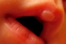
Eelt bij een pasgeboren baby: op de bovenlip, benig
Laatst beoordeeld: 07.06.2024

Alle iLive-inhoud wordt medisch beoordeeld of gecontroleerd op feiten om zo veel mogelijk feitelijke nauwkeurigheid te waarborgen.
We hebben strikte richtlijnen voor sourcing en koppelen alleen aan gerenommeerde mediasites, academische onderzoeksinstellingen en, waar mogelijk, medisch getoetste onderzoeken. Merk op dat de nummers tussen haakjes ([1], [2], etc.) klikbare links naar deze studies zijn.
Als u van mening bent dat onze inhoud onjuist, verouderd of anderszins twijfelachtig is, selecteert u deze en drukt u op Ctrl + Enter.
In kindergeneeskunde wordt een pasgeborene als een baby beschouwd binnen vier weken na de geboorte, en in deze korte tijd kan een pasgeboren blister verschijnen: en niet alleen op de lip, maar ook een botblaar.
Callus in een pasgeborene op de lip - zuigende kussen
Veel moeders die borstvoeding geven, maken zich zorgen over het zogenaamde zuigen of melk callus op de lip van een pasgeborene tijdens het geven van borstvoeding.
Inzicht in de oorzaak van het uiterlijk op de bovenlip van je baby kan hun angst elimineren.
Van de meer dan zeven dozijn aangeboren reflexen die aanwezig zijn in pasgeborenen, is een van de belangrijkste reflexen de zuigende reflex en de belangrijkste oorzaak van blaren op de bovenlip, soms in de vorm van een blaar - herhaald krachtig zuigen van melk uit de borst of uit een fles.
Bij pasgeboren baby's heeft de mondholte bepaalde functies die de baby helpen eten te "krijgen". Zuigen tijdens borstvoeding, evenals tijdens het voeden met aangepaste formulemelk, treedt op met behulp van bewegingen van de kaak en tong. En het begint met de compressie van de tepel (of facifier) door de lippen van het kind - vanwege een sterke samentrekking van de cirkelvormige spieren van de mond (musculus orbicularis oris) in de lippen en de beweging van de kauwen (musculusmasseter) van de onderste kaak, die het in het anteroposterior-vliegtuig beweegt. Deze compressie creëert de verhoogde druk die nodig is voor melkzuiging over de tepel. De baby knijpt dan dynamisch melk uit de borst in de mond door de tepel met de tong naar het harde gehemelte te knijpen.
Op dit moment is de druk in de mond lager, die niet alleen wordt geleverd door de compressie van de lippen (Musculus labii proprius Krause), maar ook door de sluiting van de interne neuspassages door het zachte gehemelte en het verlagen van de onderkaak.
Bovendien is de binnenzone van de rode rand van de bovenlip van pasgeborenen groter dan die van de onderlip en heeft een dikker en hoger epitheel met papillen - villous epitheel (waaronder een laag los bindweefsel is). Dit veroorzaakt de vorming van de pars villosa aan de grens met het slijmvliesepitheel van de lip, dat het kind helpt de tepel te begrijpen en vast te houden.
Zoals opgemerkt door neonatologen, kan de ontwikkeling van de mediale knobbeltje van de bovenlip na 9-10 weken zwangerschap in de foetus optreden (wanneer het nog steeds in de baarmoeder begint te zuigen), en in de pasgeborene is het uiterlijk van een afgeronde bulling tot 5 mm groot. En deze hobbel, hoewel het een normale anatomische variant is, wordt meestal een callus genoemd en slechts zelden als een zuigkussen. De callus kan permanent zijn, maar bij sommige baby's wordt het minder uitgesproken 10-15 minuten na het einde van elke voeding.
Het is waar dat intensief zuigen kan leiden tot de vorming van een bulla (bel) met sereuze transparante vloeistof op deze bult, en de bubbel kan barsten. Genezing vindt echter spontaan plaats - zonder behandeling - als gevolg van snelle re-epithelialisatie.
Callus op de lip van een pasgeboren baby ongemak veroorzaakt hem geen ongemak en vereist geen therapie: na een paar maanden verdwijnt het op zichzelf.
Een bot callus in een pasgeborene is het resultaat van een breuk
Het wordt algemeen erkend: in een pasgeboren babybot-callus verschijnt als gevolg van geboortetrauma, in de eerste plaats, een breuk van het sleutelbeenbot, hoewel er fracturen van andere lokalisaties kunnen zijn: humerus en zelfs dijbeen, tijdens de genezing waarvan een nieuw tissue wordt gevormd-Bone Callus in een pasgeborene.
Risicofactoren voor breuk zijn onder meer: schouderdystocie tijdens vaginale bevalling - waardoor het voor de verloskundige moeilijk is om de schoudergordel te verwijderen; belemmerde arbeid; en stuitligging van de foetus (het vergroten van de waarschijnlijkheid van dijbeenfractuur).
Buitenlandse statistieken stellen dat sleutelbeenfracturen optreden bij ongeveer een op de 50-60 pasgeborenen; Andere gegevens suggereren dat dit letsel optreedt bij ten minste 3% van de fysiologische geboorten.
Op hun beurt hebben verloskundigen een verhoogd risico op schouderdystocie (en sleutelbeenfractuur) opgemerkt in gevallen van hoog geboortegewicht - foetale macrosomie (≥4500-5000 g); In gevallen waarin een vacuüm of tang wordt gebruikt bij arbeid; zwangerschapsdiabetes (diabetische moeders hebben bredere schouders, borstomtrek en buikomtrek); Herhaalgeboorten - schouderdystocie van de pasgeborene tijdens de eerste bevalling (recidiefpercentage van dystocie wordt geschat op bijna 10%).
Daarom komt het vaker voor dat een bot callus zich vormt na een sleutelbeenbreuk in een pasgeborene.
Bij het overwegen van de pathogenese van neonatale clavicakfractuur, benadrukken experts dat het proces van ossificatie (ossificatie) van het buisvormige sleutelbeenbot (clavicula)-van de epifyseal-plaat in het centrale deel-begint in de embyo in de vijfde week van de in de vijftiende intrekerijnontwikkeling. Het mediale deel van het sleutelbeen is het dunste en de groeiplaat is open bij de geboorte, wat betekent dat het bot veel gemakkelijker te beschadigen is.
Bovendien zijn dergelijke fracturen bij pasgeborenen subperiostale fracturen, waarbij het periosteum niet wordt gestoord en de botten zelf nog steeds zacht zijn en vaak in het beschadigde deel buigen zonder uitgesproken vervorming. Fracturen van jonge zachte botten worden groene stickfracturen genoemd door chirurgen. In dit geval begint de vorming van subperiostaal nieuw bot- en bot-callus binnen tien tot tien dagen na de breuk.
De meest voorkomende symptomen van een breuk zijn lokale zwelling, rood worden van de huid, hematoomvorming, huilen van het kind bij het verplaatsen van het ipsilaterale bovenste extremiteit of gebrek aan beweging. Dit wordt pseudoparalyse genoemd: de baby stopt gewoon met het bewegen van de arm vanwege de pijn.
Gevolgen en complicaties van een dergelijke breuk zijn zeer zeldzaam: als het gebied van het letsel de groeiplaat van het bot raakt (Salter-Harris-fracturen), en een lateel wordt gevormd op de plaats van de breuk, waardoor de groei van het bot wordt vertraagd, of het is gebogen.
Diagnose bestaat uit onderzoek van de pasgeborene door een kinderarts-neonatoloog - met palpatie van de sleutelbeen, waarin de aanwezigheid van crunching reden geeft om een claviculaire breuk te diagnosticeren. Het kind wordt ook gecontroleerd op de aanwezigheid van de Moreau-reflex, en als het unilateraal (asymmetrisch) is, wordt de diagnose van breuk bevestigd.
In twijfelachtige gevallen kunnen instrumentale diagnostiek - echografie van het sleutelbeengebied - worden gebruikt. Klinische praktijk toont aan dat in sommige gevallen het sleutelbeenletsel zo onbeduidend is dat het alleen wordt gediagnosticeerd wanneer de bot callus zich begint te vormen in een pasgeborene, met het uiterlijk van een kleine uitstulping (bult) op het sleutelbeen, wat een teken is van breukverdeling.
Er wordt ook een differentiële diagnose uitgevoerd: medische professionals kunnen in een pasgeborene een zeldzame genetische botziekte detecteren - osteogenese imperfecta, myotone dystrofie of meerdere gewrichtscontracturen - arthrogryposis.
Welke behandeling is nodig als een pasgeborene een sleutelbeenbreuk heeft? Bijna al dergelijke fracturen - vanwege het grote regeneratieve potentieel van het periosteum - genezen goed zonder therapie als zodanig. Maar het is noodzakelijk om de druk en beweging van de arm van het kind aan de zijkant van het gebroken sleutelbeen te minimaliseren: immobilisatie wordt uitgevoerd door een mouw van kleding aan de zijkant van de breuk in het voorste gedeelte te bevestigen, met de arm van de baby gebogen bij de elleboog en de schouder en onderarm die aan de torso worden bevestigd. Als het huilen ernstig is, kan de arts een verdoving voorschrijven, zie voor meer informatie. - rectale pijnstillers en ontstekingsremmende zetpillen.
Het is normaal dat een kind na ongeveer twee weken de arm aan de zijkant van de breuk gaat verplaatsen.
Zoals de onderzoekers ontdekten, ontstaat de zachte callus op de breuklocatie uit kraakbeen en creëert, door aan één kant van de breuk te groeien een kracht die het beschadigde bot uitlijnt. De verharding van de callus bevordert de volledige genezing van de breuk en duurt gemiddeld vier tot vijf weken.
De preventie van schouderdystocie die door sommige clinici wordt aanbevolen, is een keuzevak een keizersnede voor zwangere vrouwen wier pasgeborene een geschiedenis heeft van een sleutelbeenbreuk. Maar experts van het American College of Obstetricians en Gynaecologen (ACOG) beschouwen het voordeel van een dergelijke preventieve maatregel.
Bovendien brengt een keizersnede in noodsituaties een hoger risico op een lange botbreuk met zich mee dan een normale afgifte.
Zoveel experts zijn geneigd te geloven dat een neonatale sleutelbeenbreuk tijdens de bevalling waarschijnlijk niet te voorkomen is.
De prognose voor een sleutelbeenfractuur tijdens de bevalling is echter uitstekend, en de bot callus in een pasgeborene nadat een sleutelbeenbreuk binnen zes maanden verdwijnt.